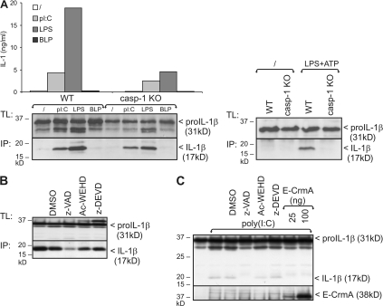
Figure 3.
Poly(I:C) and LPS-induced pro–IL-1β processing is caspase-1 independent. (A) BLP-primed peritoneal macrophages from WT and caspase-1 KO mice were incubated for 18 h with poly(I:C), LPS, BLP, or control medium, as described in the Materials and methods. Pro–IL-1β processing was analyzed by Western blotting of total cell lysates (TL) and IL-1β immunoprecipitates (IP) from the cell supernatant (left). As a positive control for caspase-1–mediated pro–IL-1β processing, cells were stimulated for 12 h with 100 ng/ml LPS, pulsed for 20 min with 5 mM ATP, and subsequently incubated in fresh medium for 3 h (right). (B) Peritoneal macrophages were stimulated for 18 h with poly(I:C), as described in A. 1 h before incubation, cells received 50 μM z-VAD-fmk, Ac-WEHD-cho, z-DEVD-cmk, or 0.05% DMSO (solvent control). Pro–IL-1β processing was analyzed by Western blotting of total cell lysates (TL) and IL-1β immunoprecipitates (IP) from the cell supernatant (bottom). (C) HEK293-TLR3 cells transfected with 0.6 μg pCAGGS-pro–IL-1β were incubated for 6 h with 25 μg/ml poly(I:C). 1 h before incubation, cells received 50 μM z-VAD-fmk, Ac-WEHD-cho, z-DEVD-cmk, or 0.05% DMSO (solvent control). In the last two lanes, HEK293-TLR3 cells were cotransfected with two different concentrations of CrmA-E. Pro–IL-1β processing was analyzed by SDS-PAGE and Western blotting of total cell lysates. Expression of CrmA was verified by Western blotting and anti-E tag. Data are representative of three independent experiments.

