Figure 2.
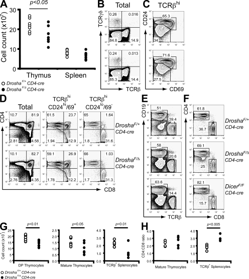
Drosha deficiency at the DP thymocyte stage partially perturbs thymocyte output. (A) Total thymocyte and splenocyte numbers in DroshaF/Δ CD4-cre and DroshaF/+ CD4-cre (control) mice. (B) TCRβ versus TCRγδ expression on total thymocytes. (C) CD24 and CD69 expression on TCRβhi gated thymocytes. (D) CD4 versus CD8 expression on total, postselected (TCRβhiCD24hi/69+) and mature (TCRβhiCD24lo/69−) thymocytes. (E and F) FACS analysis of populations in total splenocytes (E) and TCRβ+ splenocytes (F). Also shown is the effect of Dicer deficiency. Percentages of cells are shown in B–F. (G) Absolute CD4+8+ DP, mature thymocyte, and TCRβ+ splenocyte numbers. (H) CD4/CD8 ratio in the mature thymocyte and TCRβ+ splenocyte compartments.

