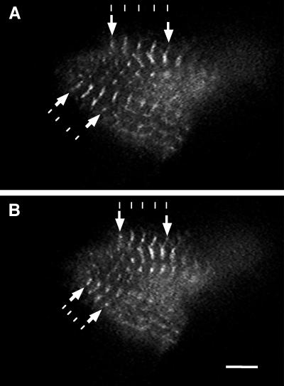
Figure 4.
Living cardiomyocyte expressing cMy2–GFP. Single images taken from a video recording of a cardiomyocyte expressing cMy2–GFP showing relaxed (A) and contracted (B) states. cMy2–GFP localizes to the M-bands of the sarcomere and does not inhibit the contractile activity of the cardiomyocyte. The contraction of the sarcomeres can be seen by comparing the decrease in distance between neighboring M-bands in the contracted and uncontracted state (arrows). The lines are drawn to visualize the shortening of the distance between neighboring M-bands. Bar, 5 μm.

