Figure 3.
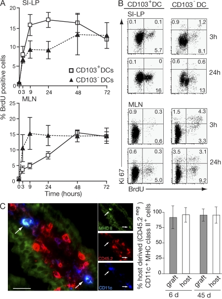
Turnover of SI-LP and MLN CD103+ and CD103− DC. (A) Mice were injected i.p. with 2 mg BrdU, and the percentage of BrdU+ CD103+ and CD103− DCs (MHC class II+CD11c+) in the SI-LP and MLN was determined by flow cytometry at the times indicated. Results are the mean and SD of three to seven independent experiments with two mice in each time point except the 72-h time point, which was performed once. (B) BrdU and Ki67 staining on SI-LP and MLN CD103+ and CD103− DCs (MHC class II+ CD11c+) was assessed by flow cytometry 3 and 24 h after BrdU injection. Plots are from one representative experiment of three performed. (C) SI from CD45.2+ mice (graft) was transplanted into CD45.1+ recipients (host) as previously described (32). At days 6 and 45, host and graft intestine were sectioned and stained with antibodies to CD45.2 (red), CD11c (blue), and MHC class II (green). Immunohistochemistry of the 45-d graft is shown. Arrows point to host-derived CD45.2−MHC class IIhiCD11c+ DCs. Bar, 25 μm. The graph is percentage of host-derived (CD45.2 negative) CD11c+MHC class IIhi cells in graft and host small intestinal villus. Results are mean and SD (n = 3 mice per group).

