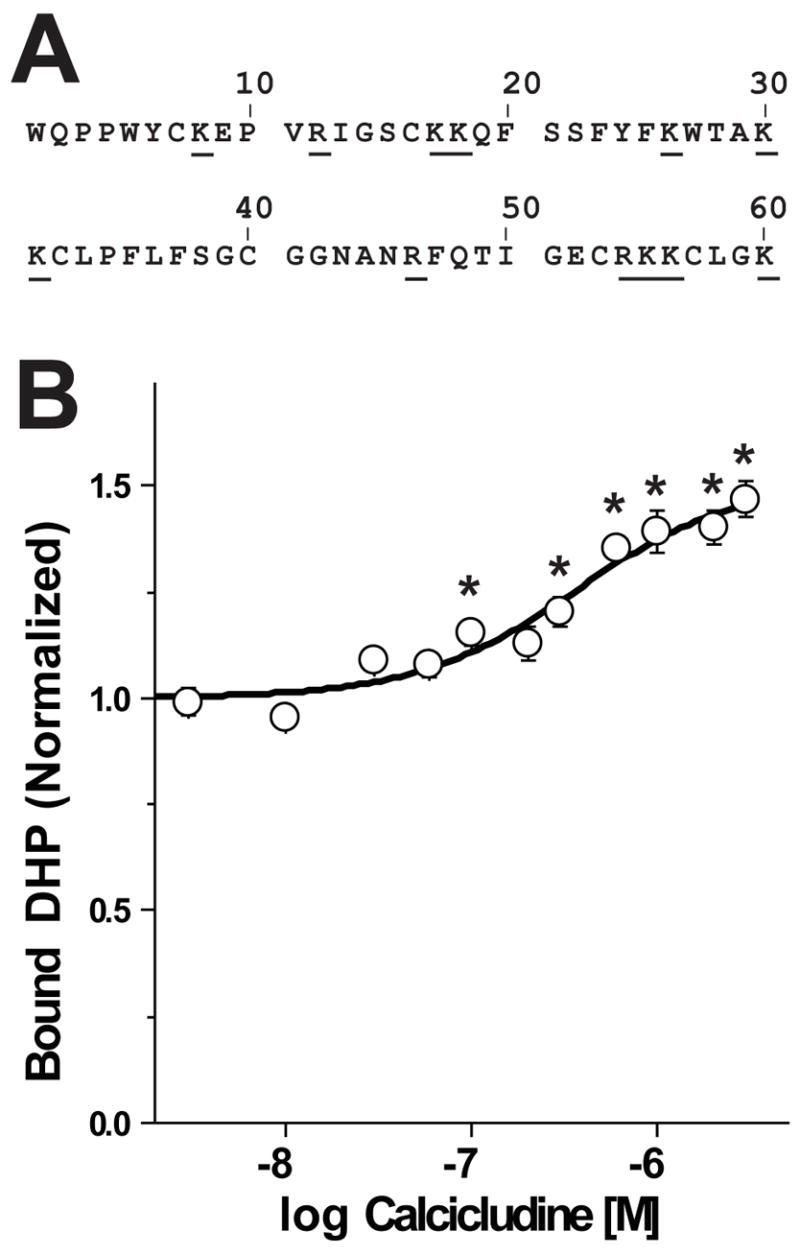
FIGURE 1. Calcicludine is a positive allosteric modulator of DHP binding. (A) Primary sequence of calcicludine.

Calcicludine is a 60 amino acid peptide containing three disulfide bonds, 12 positively charged lysine or arginine residues (underlined) and two negatively charged glutamate residues. (B) Membranes from cells expressing wild-type CaV1.2 Ca2+ channels were incubated with ~350 pM [3H]PN200-110 and increasing concentrations of calcicludine. Each experiment was fit using Scheme 1 (see Equation 4, Materials and Methods) and normalized such that the occupancy in zero calcicludine is equal to 1.0. Note that [3H]PN200-110 binding increases as calcicludine is raised from 3 nM to 3 μM. Binding data are means ± SEM, and calcicludine concentrations where DHP binding differs significantly from that with no toxin (determined by ANOVA) are indicated with asterisks (*; P<0.05; n=6). Error bars smaller than symbols do not appear in figures. (C) Allosteric binding model for DHP and calcicludine binding. See Materials and Methods and text for details.
