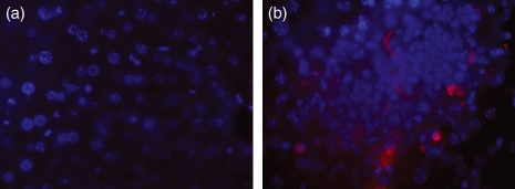
Figure 2.
Multicolour fluorescence microscopy showing (a) the absence of caspase-3-positive cells in uninfected tissue, (b) a caspase-3-positive cell within a focus of infection 96 hr postinfection in BALB/c mice infected with Salmonella Typhimurium C5. (Caspase-3-positive cells were detected with Anti-Caspase-3 antibody/Alexa Fluor 568 and appear red, nuclei are stained with DAPI and appear blue. Images were taken at magnification × 630).

