Figure 1.
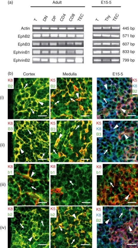
Expression of EphB receptors and ephrinB ligands in mouse thymus. (a) Specific primer pairs were used to determine by RT–PCR analysis the presence of EphB2, EphB3, ephrinB1 and ephrinB2 in total thymus (T), total thymocytes (Thy) isolated thymocyte subpopulations based on CD4, CD8, and TCRαβ expression. (DN: CD4− CD8− TCRαβ−; DP: CD4+ CD8+ TCRαβ+/−; CD4: CD4+ CD8− TCRαβ+; CD8: CD4− CD8+ TCRαβ+) and isolated thymic epithelial cells (TEC). Samples were obtained from adult or 15.5 embryonic (E15.5) WT mice. β-actin served as positive control. Corresponding band sizes are indicated on the right side of the figure. (b) Topological detection of Eph/ephrinB on thymus cryosections from adult and 15.5 embryonic (E15.5) WT mice. EphB2 (i), EphB3 (ii), ephrinB1 (b1) (iii) and ephrinB2 (b2) (iv) are expressed on both thymocytes (keratin negative cells; arrows) and epithelial cells (arrowheads) of thymic cortex (K8+ cells) and medulla (K5+ cells). Scale bar 10 μm.

