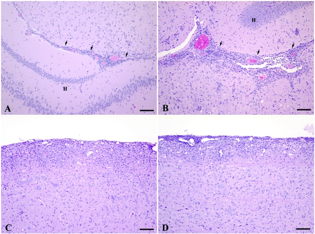
Figure 3.
Comparison of the histopathological lesions caused by EAE in the brains of WT (A) and VWFKO (B) mice and the spinal cords of WT (C) and VWFKO (D) mice. In the brains, the inflammatory response, consisting of an admixture of lymphocytes and monocytes with occasional neutrophils and rare eosinophils, was more severe in the VWFKO mice (B) than the response that occurred in the brains of the WT mice (A). In the brains of both strains of mice, the inflammatory response commonly occurred around capillaries and postcapillary venules in the interface area (arrows) between the brainstem and hippocampal formation (H). Mild demyelination occurred concurrently with inflammation in the VWFKO strain of mice; however, none was observed in the WT strain of mice. A and B: H&E stain, scale bar = 100 μm. In the spinal cords, the severity and character of the inflammatory response were similar between WT (C) and VWFKO (D) mice and again consisted of an admixture of lymphocytes and monocytes with occasional neutrophils and rare eosinophils. The inflammatory response commonly occurred around capillaries and post capillary venules in the pia-arachnoid and subjacent white matter. Mild demyelination occurred concurrently with inflammation in both strains of mice. C and D: H&E stain, scale bar = 100 μm.

