Figure 8.
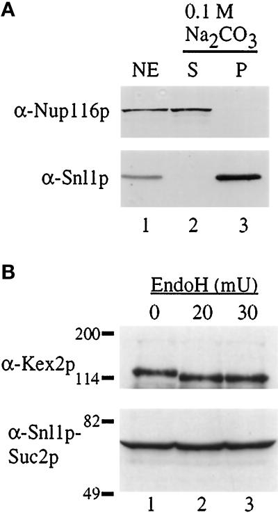
Snl1p is an integral membrane protein, oriented with the C-terminal region accessible to the cytoplasm. (A) Purified nuclear envelopes (NE, lane 1) from W303 diploid nuclei were treated with 0.1 M Na2CO3 (pH 11) and separated into supernatant (S, lane 2) and pellet (P, lane 3) fractions by centrifugation. Samples were separated on a 13% SDS-polyacrylamide gel, transferred to nitrocellulose, and immunoblotted. Strips corresponding to the molecular mass of Nup116p (top) and Snl1p (bottom) were cut from the same blot and probed with the respective antibodies. (B) Cell extracts from a strain expressing an Snl1p–Suc2p fusion protein were either mock treated (0, lane 1) or treated with EndoH (lane 2, 3) as described in MATERIALS AND METHODS (mU = milliunits). The samples were TCA precipitated and resuspended in SDS sample buffer for separation on a 7% SDS-polyacrylamide gel. Immunoblot analysis was performed for Kex2p (top) and Snl1p–Suc2p (bottom). Molecular mass markers are indicated in kDa.

