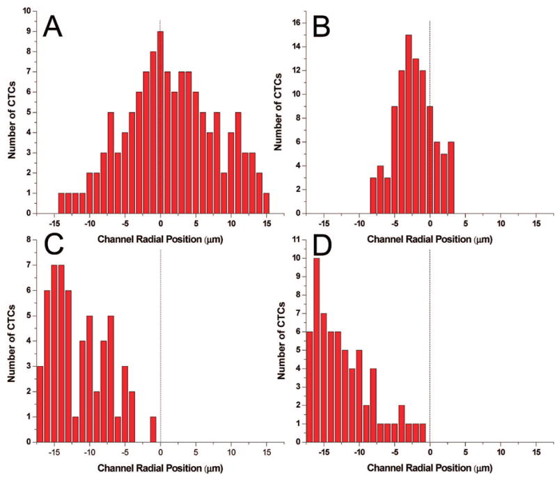
Figure 2.

Histograms of the radial position of CTCs (centroid) in microchannels with Poiseulle flow at linear velocities (U) of 1.0 and 10 mm s−1 in both straight (A, B) and sinusoidal-configured (C, D) channels. The dashed line represents the microchannel’s central axis. (A and B) The radial position of several CTCs histogrammed from micrographs of the straight microchannel with linear flow rates of 1 and 10 mm s−1. (C and D) The radial position of several CTCs traversing 1/4 of a period of the sinusoidal microchannels with suspension linear velocities of 1 and 10 mm s−1 are shown. The cells were imaged using fluorescence microscopy with the cells stained using a fluorescein lipophilic membrane dye, PKH67.
