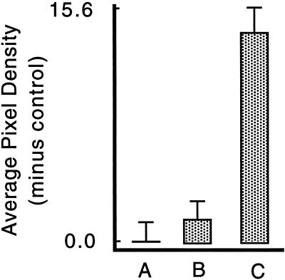
Figure 6.
Fos expression is inhibited by elevated extracellular Ca2+. BAE cell monolayers were washed into PBS saline (A, C), or iso-osmolar NaCl, HEPES-buffered saline containing 50 mM Ca2+ (B), both of which contained FDx. Five minutes later, all except two control PBS-washed cultures (A) were scratched in these salines, and the fos expression level was quantitated in every cell encountered along scratch zones or at random in the undisturbed cultures (A). The high calcium treatment eliminated FDx-positive cells along the scratch zone (our unpublished results) and hence selectively eliminated PMD-affected cells from the responding monolayer. Therefore, unlike in other experiments, we could not correlate FDx and c-fos intensities in this analysis because the cultures injured in high Ca2+ entirely lack FDx-positive cells. The values shown represent the mean and SEs of the fos intensity measured by image analysis from ∼150 cells on replicate coverslips after subtraction of the mean value measured from an equivalent number of control, undisturbed cells (A), which represents “autofluorescence” background. The fos response of the elevated Ca2+ cultures (B) was not significantly different from the control, undisturbed culture (A) (p = 0.3993), but was significantly reduced relative to the PBS (C) cultures (p < 0.0001).

