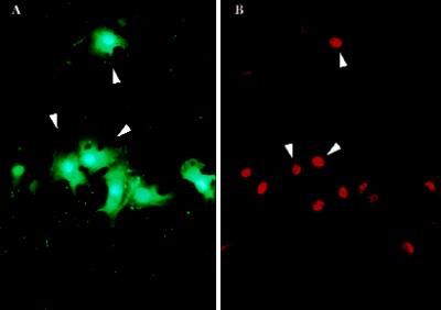
Figure 8.
NF-κB translocation into the nucleus is also induced along monolayer injury sites where PMD is common. (A) A confluent monolayer of BAE cells was scratched in FDx. As in Figure 1, cells incurring a survivable PMD are labeled with this marker. (B) A paired image showing immunostaining for NF-κB. Note the correspondence of PMD and NF-κB events, as was the case for the fos event.

