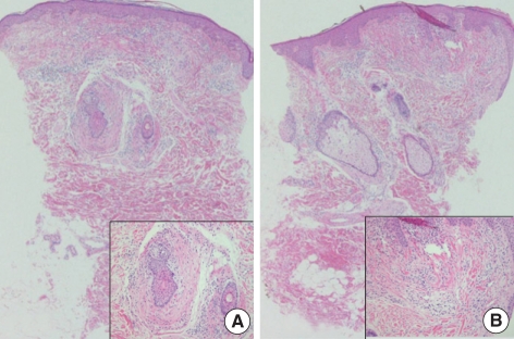
Fig. 2.
(A) Concentric layers of cellular fibrous tissue around hair follicles representing perifollicular fibroma (hematoxylin-eosin stain; original magnification ×40, inset ×200). (B) Raised papule composed of connective tissue surrounded by a lateral collarette representing trichodiscoma (hematoxylin-eosin stain; original magnification ×40, inset ×200).

