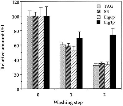
Figure 3.
Presence of Erg1p in the endoplasmic reticulum was not due to a contamination with lipid particles. Endoplasmic reticulum (30,000 × g microsomes) was prepared as described in MATERIALS AND METHODS and subjected to two additional washes with 10 mM Tris-HCl, pH 7.5. Triacylglycerols (TAG) and steryl esters (SE) were quantified after thin-layer chromatographic separation of lipids (see MATERIALS AND METHODS), and Erg6p and Erg1p in the membrane pellet were quantified before and after the respective washing steps by Western blot analysis. Mean values of three independent experiments are shown.

