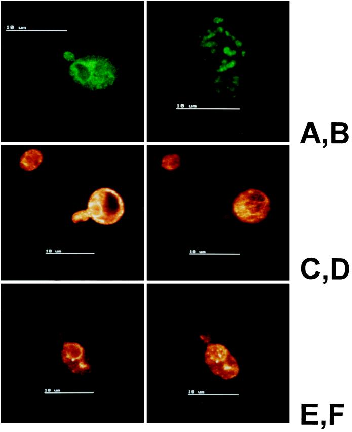
Figure 4.
Indirect immunofluorescence microscopy of Erg1p in yeast at various growth stages. Cells of the diploid strain Saccharomyces cerevisiae W303D were prepared as described in MATERIALS AND METHODS, and immunoreactive proteins were detected with FITC-conjugated anti-rabbit IgG. Primary antibodies against the endoplasmic reticulum marker BiP (Kar2p) (A), the lipid particle marker sterol Δ24-methyltransferase (Erg6p) (B), and Erg1p (C—F) were used. (C and D) Localization of Erg1p in two sections of the same cell in the logarithmic phase. (E) Localization of Erg1p in a single optical section of a late logarithmic-phase cell. (F) Extended focus image of the same cell.

