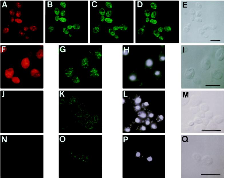
Figure 5.
Localization of Erg1p in the endoplasmic reticulum and a subpopulation of lipid particles. Late-logarithmic cells of a tetraploid wild-type strain (A–I) and the erg1 disruptant strain KLN (J–Q) were prepared for double immunofluorescence microscopy as described in MATERIALS AND METHODS. (A–E) Localization of Erg1p (A) and BiP (B–D; three different optical sections) in the same wild-type cells. (E) Transmission image. (F–I) Localization of Erg1p (F) and Erg6p (G) in the same wild-type cells. (H) DAPI staining. (I) Transmission image. (J–Q) erg1 disruptant strain KLN lacks a distinct signal with Cy5-labeled Erg1p antibody (J and N). Localization of BiP (K) and Erg6p (O), with DAPI staining (L and P) and transmission images (M and Q) of the respective cells of the strain KLN. Bar, 10 μm.

