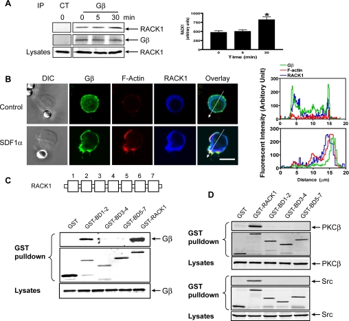
Figure 3.
Interaction of RACK1 with Gβγ. (A) Association of RACK1 with Gβγ was determined after stimulation of Jurkat cells with SDF1α (20 nM) for the indicated times, and Gβγ was immunoprecipitated from the cell lysates with a control antibody (CT) or Gβ antibody that recognizes Gβ1, 2, 3, and 4 isoforms. The presence of RACK1 and Gβ in the precipitate was detected by specific antibodies. Left panel, representative Western blots; right panel, quantitative data. *p < 0.05, significant difference versus 0 min. (B) Colocalization of RACK1 and Gβγ in Jurkat cells. Cells were treated with control beads or SDF1α-conjugated beads for 2 min and then stained with specific antibodies or Alexa-568–conjugated phalloidin for F-actin. Distraction interference contrast (DIC) and fluorescent images are shown. Bar, 10 μm. The graphs on the right panel show the distribution of fluorescent intensity of Gβ, F-actin, and RACK1 along the line drawn across the cells. The images are the representatives of more than 20 cells from at least three separate experiments with similar results. (C and D) Association of RACK1 and its mutants with Gβ1γ2 (C), PKCβ or Src (D) was determined by GST pulldown assays after coexpression of HA-Gβ1γ2, PKCβ, or Src with the indicated constructs in HEK293 cells. The presence of the proteins in the GST pulldown pellets and lysates was detected with specific antibodies. A schematic representation of RACK1 structure is shown in the top panel of C.

