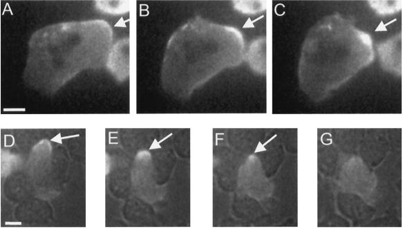
Figure 2.
Examples of the C-to-spot transition during pseudopod retraction and cell squeezing. (A–C) C condensing to a spot (arrows) in a pseudopod as it retracts in a starved GFP-MHC cell resting on a substrate. (Also see online movies, or the author’s web site listed in ACKNOWLEDGMENTS.) (A) C, 0 s; (B) condensing C, 35 s; (C) spot, 65 s. (A second pseudopod in the same cell is being retracted just below the spot marked by the arrow. This second pseudopod is in the process of condensing to a spot.) (D–F) C condensing to a spot as a starved cell squeezes between two neighbors. The image was obtained by simultaneous fluorescence and bright-field microscopy. A GFP-MHC–labeled cell is moving between several unlabeled adjacent cells. Arrows indicate the C at the cell posterior (D, 0 s) undergoing condensation (E, 23 s) to a spot (F, 39 s), which eventually disintegrates (G, 62 s). Bars, 5 μm.

