Figure 7.
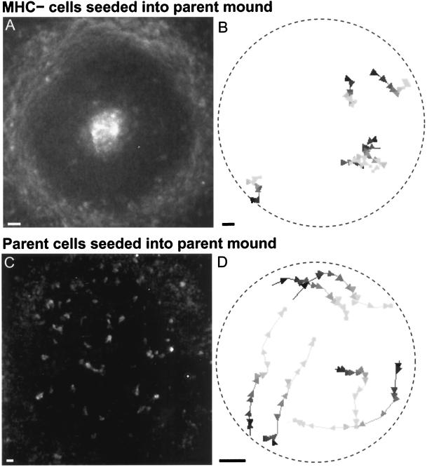
Fluorescently labeled cells seeded into a mound composed of parent strain cells. (A) In such chimeric mounds, MHC− cells segregated to the inner core or outer periphery. Shown is one image from a time-lapse movie obtained ∼3 h after a small number of starved MHC− cells were added to a mound of unlabeled parent strain cells. (B) MHC− cells fail to rotate in such chimeric mounds. Five sample trajectories are shown (the two near the 4:00 position partially overlap). Conventions for trajectory display are as described in Figure 4. Arrowheads represent time points 1 min apart. The slow, irregular movements of the MHC− cells indicate that the spiral wave generated by the parent cells is not enough to restore normal motion in MHC− cells. (C) Control. One time point of a 2D time-lapse movie taken ∼3.5 h after starved parent cells were added to a parent mound. Fluorescently labeled parent cells do not segregate in these mounds. (D) Typical trajectories of parent cells added to a parent strain mound. The seeded cells rotate normally. Arrowheads represent time points 40 s apart. Bars, 10 μm.

