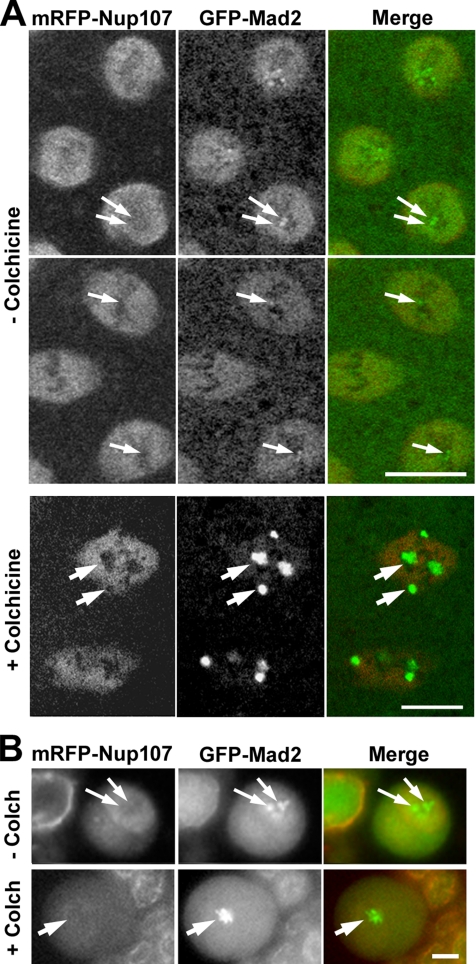
Figure 10.
Nup107 is not detectable at kinetochores during Drosophila mitosis. (A) Confocal images of syncytial stage embryos expressing mRFP-Nup107 and GFP-Mad2 either untreated (−colchicine) and recorded in prometaphase and metaphase (30 s between the two frames), or imaged ∼5–10 min after microinjection with colchicine (1 mM). (B) Wide-field images of live larval neuroblasts expressing mRFP-Nup107 and GFP-Mad2 either untreated (−Colch) or imaged after an ∼10-min incubation with colchicine (10 μM). There is no detectable mRFP-Nup107 at kinetochores (arrows) in either cell type, even upon colchicine treatment, which leads to substantial accumulation of GFP-Mad2 on these structures. Note also that colchicine treatment does not affect the accumulation of Nup107 in the spindle region (in A, +colchicine). Bars, 5 μm.

