Figure 2.
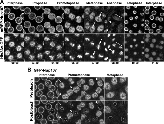
Dynamics of mRFP-Nup107 throughout the mitotic cycle in Drosophila syncytial embryos. (A) Selected frames of TLSCM acquisitions revealing the dynamics of mRFP-Nup107 together with His2Av-GFP during a complete embryonic cleavage cycle. A representative focal plane from a 3.6-μm z-stack acquisition is shown for each time point. Note the decreased NE localization and concomitant nuclear accumulation of mRFP-Nup107 in prometaphase and its persistence within the spindle area in metaphase. Arrows point to the chromatin-occupied (i.e., GFP-histone-labeled) areas from which mRFP-Nup107 is excluded. In anaphase, the signal fades within the spindle area (brackets) and redistributes over the segregated chromatids (arrowheads). The first rims reappear on the decondensing chromatin in late anaphase/early telophase. Time is in minutes:seconds. Bar, 10 μm. (B) Photobleaching analysis of GFP-Nup107 in embryonic nuclei. Small regions (outlined in white in the frames) within nuclei of GFP-Nup107 Drosophila embryos at various stages of the cell cycle were bleached. Acquisitions before and just after photobleaching are shown. Bars, 5 μm. Note the stable signal at the NE in interphase nuclei and the persistence of a minor fraction of GFP-Nup107 stably associated with the NE in early (arrowheads) but not late (arrows) prometaphase stages.

