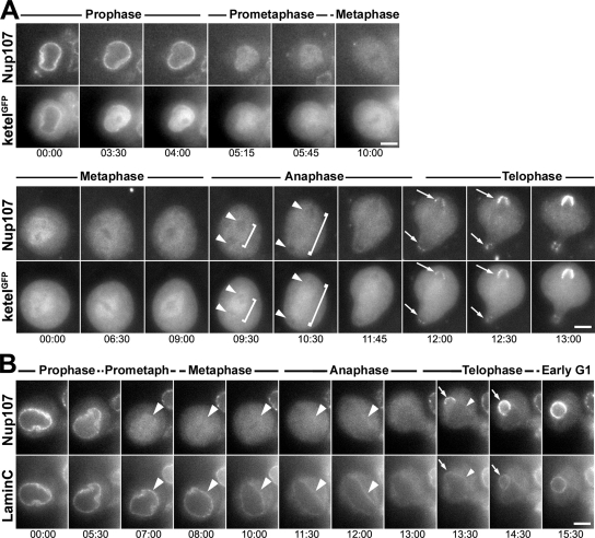
Figure 6.
NPC and NE dynamics in dividing larval neuroblasts. (A) Selected frames of wide-field time-lapse acquisition of dividing larval neuroblasts expressing mRFP-Nup107 and ketelGFP. Two representative series describing the prophase to metaphase (top) and the metaphase to telophase stages (bottom) are shown (see also Supplemental Movies 7 and 8). Note that ketelGFP is released from the prophase NE earlier than mRFP-Nup107 (top, 3:30 and 4:00 min). Both proteins are enriched within the area defined by the spindle envelope in early metaphase, and a fraction of ketelGFP but not mRFP-Nup107 persists within the spindle area in anaphase (brackets). Note that at these stages, both Nup107 and ketelGFP are always excluded from the chromatin area (arrowheads). The proteins are recruited simultaneously to the reforming nuclear envelope in early telophase (arrows). (B) Selected frames of wide-field time-lapse acquisition of a dividing larval neuroblast expressing mRFP-Nup107 and GFP-Lamin C. Note the persistence of the GFP-Lamin C staining at the spindle envelope until late anaphase (arrowheads). Arrows point to the reforming NE and NPCs in telophase. Bars, 5 μm. Time is in minutes:seconds.

