Figure 9.
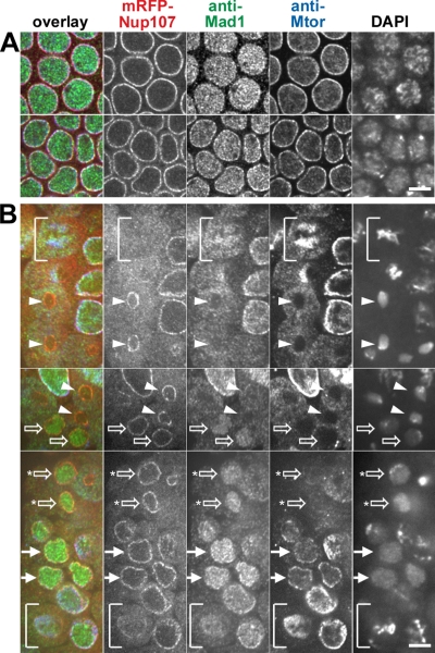
Localization of mRFP-Nup107, Mad1, and the basket nucleoporin Mtor during embryonic mitotic divisions. Spinning disk confocal images of fixed syncytial and cellularized mRFP-Nup107 Drosophila embryos labeled with anti-Mad1 (green), anti Mtor (blue), and DAPI (not included in the overlay). (A) Unlike mRFP-Nup107 and Mtor, Mad1 is mainly localized in the nucleoplasm in syncytial embryos (top), and its NE localization becomes clearly detectable in cellularized embryos (bottom). (B) Successive steps of Mad1 and Mtor recruitment in the nucleus and at the NE during mitotic divisions of a cellularized embryo. The three panels arise from distinct areas and focal planes within the same embryo. The bracket in the bottom panel points to a prometaphase cell in which a fraction of Nup107 is still detectable at the NE, whereas Mad2 and Mtor are localized within the spindle area. The bracket in the top panel shows a metaphase cell revealing the typical localization of Mtor at the spindle matrix. Note that a fraction of Mad1 also accumulates within the spindle area whereas mRFP-Nup107 is more diffuse. Arrowheads point to cells in telophase in which Mad1 is still in the cytoplasm. Open arrows indicate cells with a nuclear accumulation of Mad1 and a cytoplasmic localization of Mtor. Some Mtor begins to be detectable at the NE in cells with an asterisk. Solid arrows indicate G1 cells in which Mtor and a fraction of Mad1 are localized to the NE. Bars, 5 μm.

