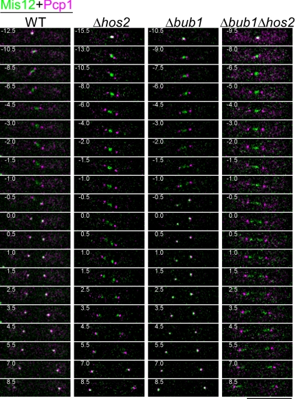
Figure 5.
Δhos2 cells are defective in poleward movement of kinetochores during anaphase A. Time-lapse images of mitosis in exponentially growing cells with the indicated genotype are presented. Kinetochores and the SPB were visualized by Mis12-GFP (green) and Pcp1-CFP (magenta), respectively. Cells were cultured in minimal medium at 26°C, and stacks of Z-sections with an interval of 0.4 μm were taken every 0.5 min. The numbers indicate the duration in minutes. The time point at which phase 3 spindle extension began was defined as 0. Bar, 10 μm.

