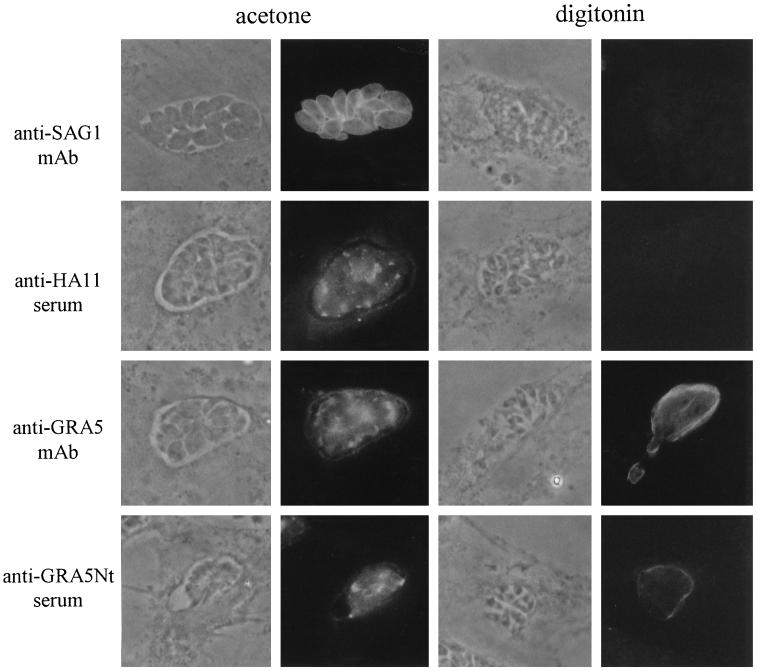
Figure 7.
Orientation of the GRA5–HA9 protein within the PVM by immunofluorescence staining. Cells are visualized by phase-contrast microscopy. Immunofluorescence experiments were performed on cells infected with the GRA5–HA9 Toxoplasma clone. Cells were permeabilized either completely by cold acetone (left) or selectively by 0.004% digitonin (right). Left, when cells were permeabilized by acetone, the parasite surface was stained by the anti-SAG1 mAb. Under these conditions, the staining patterns obtained with both anti-GRA5 antibodies (the TG17–113 mAb and the anti-GRA5 Nt serum) and the anti-HA11 rabbit serum were identical; GRA5–HA9 was detected associated with the PVM and in clusters between the parasites. Right, when the host cell membrane was selectively permeabilized by digitonin, as verified by the negative staining of the parasite surface with the anti-SAG1 mAb, the C-terminal part of GRA5–HA9, recognized by the anti-HA11 serum, was not detected. In contrast, the external side of the PV was stained by both the anti-GRA5 Nt serum and the anti-GRA5 mAb.

