Figure 20.

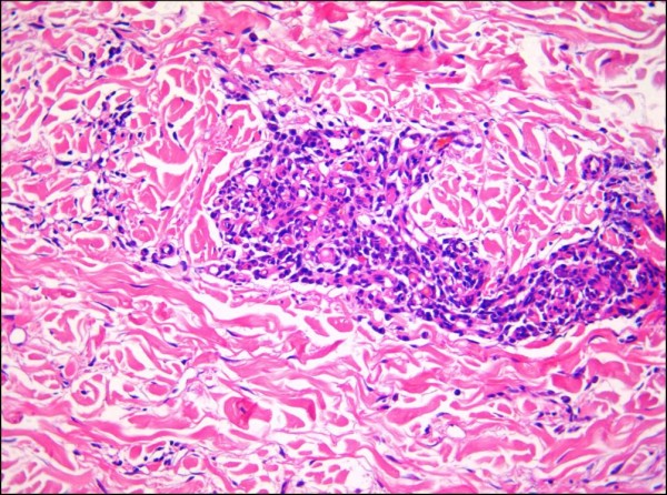
Regressed Kaposi sarcoma lesion seen at higher magnification showing a perivascular infiltrate comprised mainly of plasma cells (H&E stain).

Regressed Kaposi sarcoma lesion seen at higher magnification showing a perivascular infiltrate comprised mainly of plasma cells (H&E stain).