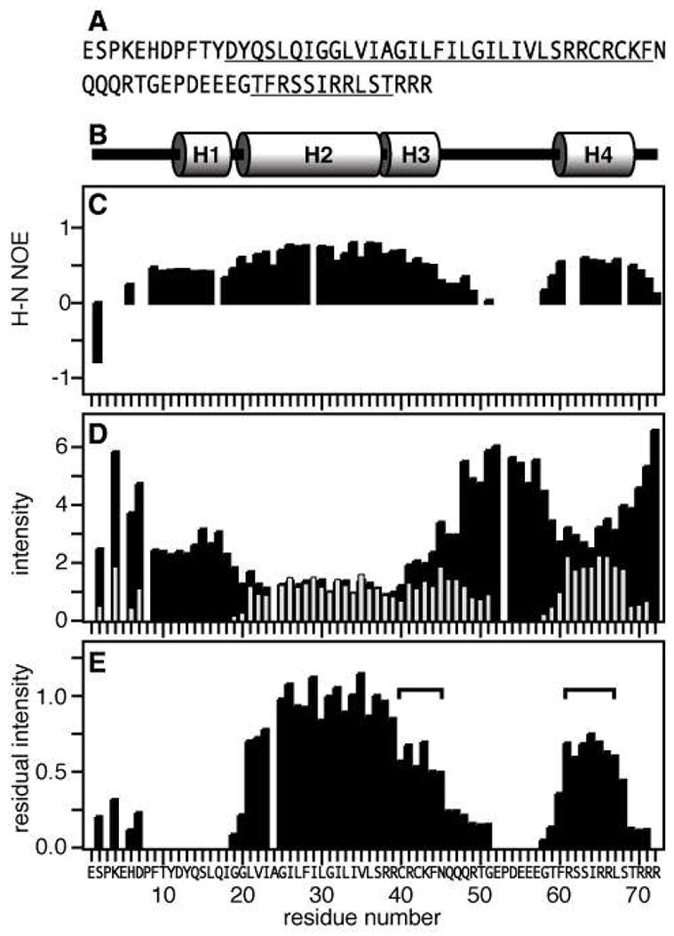
Figure 2.

Effect of Mn-induced PRE on the resonance intensities. (A) Amino acid sequence of human FXYD1 with helical regions underlined. Residue numbering begins at 1 after the signal sequence (NCBI protein accession: NP_068702). (B) Protein secondary structure. (C) 1H/15N heteronuclear NOEs. (D) Normalized 1H/15N HSQC peak intensities obtained without (I−Mn, black bars) or with (I+Mn, gray bars) 1.6 mM MnCl2. Positions that are left blank correspond to prolines (P3, P8, P53) or overlapped resonances (E5, A24). (E) Residual normalized peak intensity (I−Mn/I+Mn). The horizontal square brackets mark residues in H3 and H4 with similar protection from aqueous Mn.
