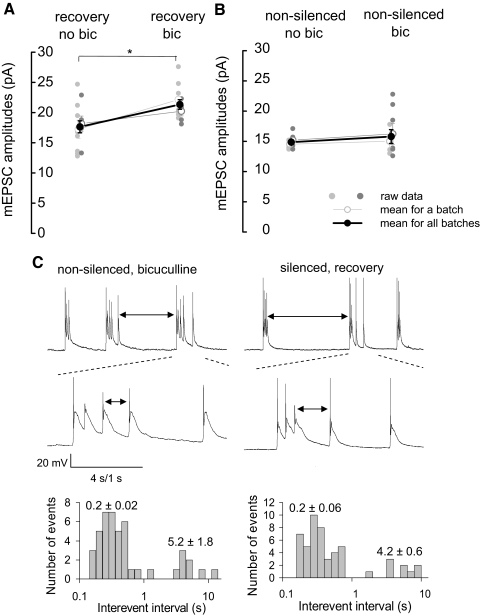
FIG. 3.
Similar bursting has different outcome on mEPSC potentiation in silenced and nonsilenced neurons. A: mEPSC potentiation in silenced cultures following a 20-min recovery of activity in 20 μM bicuculline-containing bathing solution was more pronounced than that in bicuculline-free solution. mEPSC amplitudes were significantly higher (21 ± 4%) in cultures recovering in the presence of bicuculline than those without. *, significance in the statistical data analysis (2-way ANOVA, P < 0.05). B: disinhibition of control (nonsilenced) neurons with bicuculline (20 μM) for 20- 25 min did not significantly change mEPSC amplitudes. In A and B, data from 2 batches of neurons are presented (small gray circles). The average for each batch is presented as open circles. The average mEPSC amplitudes for the 2 batches of cultures are presented as black circles (n = 4–8 neurons/batch). C, top: representative traces of membrane potential recordings in nonsilenced neurons disinhibited by bicuculline (left) and in silenced neurons during the recovery period (right). In both groups of neurons, firing of action potentials occurred in bursts with similar patterns. ↓, interburst intervals. Middle: expanded view of the burst of action potentials of nonsilenced neurons disinhibited by bicuculline (left) and in silenced neurons during the recovery period (right). ↓, intraburst intervals. Bottom: histograms of log-binned (10 bins/decade) action potential interspike intervals in 2 recordings. Values represent the average intraburst interspike intervals (0.2 ± 0.02 and 0.2 ± 0.06 s for nonsilenced and silenced neurons, respectively), and the average interburst intervals (5.2 ± 1.8 and 4.6 ± 0.6 s for nonsilenced neurons and silenced neurons, respectively).

