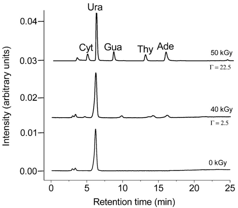
FIG. 1.

HPLC chromatograms (monitored at 254 nm) are shown for pUC18 films incubated at Γ = 2.5 and 22.5 and X-irradiated with 40 kGy and 50 kGy, respectively. For a dose of 0 kGy, a Γ = 2.5 film is shown; the Γ = 22.5 is effectively the same. Cyt = cytosine, Gua = guanine, Ade = adenine, Thy = thymine, and Ura = uracil; the latter was used as an internal standard. Note that in comparing peak intensities, it is important to take into account the difference in target mass between Γ = 2.5 and Γ = 22.5.
