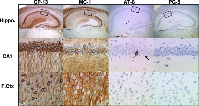
Figure 4.
Accumulation of pathological tau species in rTg3696AB brain. At 4 months of age, IHC studies revealed abnormal tau conformation and phosphorylation in rTg3696AB hippocampus and frontal cortex. Enlarged images of hippocampal subdivision CA1 are shown in middle panel. At this age, neurons were positively labeled using antibodies directed at pathological epitopes dependent on changes in conformation (MC-1, amino acids 7 to 9 and amino acids 326 to 300) and phosphorylation (CP-13, pSer202; AT-8, pSer202/pThr205; PG-5, pSer409). Boxes indicate area shown at higher magnification. Rare AT-8-positive CA1 neuron indicated by arrow. Original magnifications: ×5 (top); ×40 (middle, bottom).

