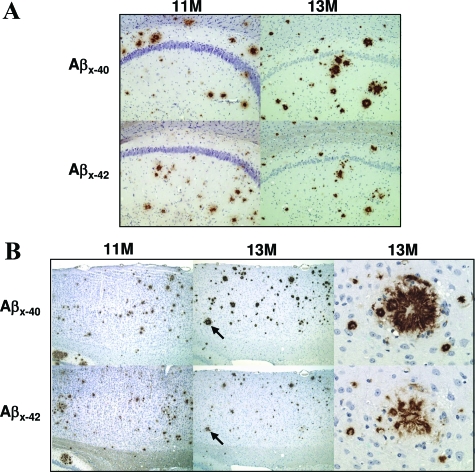
Figure 5.
Age-dependent progression of plaque pathology. IHC images indicate substantial accumulation of Aβ plaque pathology in rTg3696AB mice between 11 and 13 months of age. Representative photomicrographs depict hippocampal (A) and cortical (B) pathology detected with Aβx-40 and Aβx-42. Arrows indicate specific plaques shown at high magnification. Original magnifications: ×10 (A); ×5 (B, low-power cortical images); ×40 (B, high-power plaque images).

