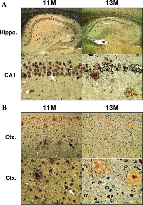
Figure 7.
Progression of mature plaque and NFT pathology. Tissue was impregnated with Bielschowsky silver stain to visualize mature plaque and tangle pathology. A: Representative images are shown for hippocampus, including higher magnification of CA1 (bottom). B: NFT pathology was also evident in frontal cortex. NFT pathology can most clearly be observed in high-power images (bottom). Rare Bielschowsky-positive NFTs in 11-month brain highlighted with arrow. Boxes indicate area shown at higher magnification. Original magnifications: ×5 (A, top); ×10 (B, top); ×40 (A, B, bottom).

