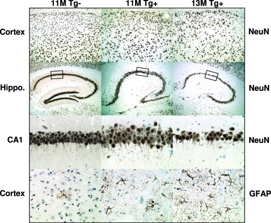
Figure 9.
Neuronal loss and astrogliosis in aged rTg3696AB mice. Tissue was processed with neuron-specific antibody NeuN. Images indicate substantial loss of neurons in rTg3696AB mice between 11 and 13 months. Representative photomicrographs depict cortex (top), hippocampus, and CA1 hippocampal subdivision (middle) after processing with NeuN. In parallel, GFAP was used to detect glia cells (bottom). Original magnifications: ×10 (top); ×5 (top middle); ×40 (bottom middle and bottom).

