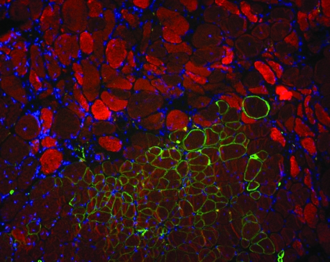
Figure 3.
AMMC-derived muscle fibers resist degeneration. For each recipient, the serial tibialis anterior section containing the most donor-derived myofibers was stained with anti-γ-sarcoglycan antibodies (green) and EBD (red), which binds albumin and enters damaged muscle fibers. Nuclei are stained with DAPI (blue). Damaged muscle fibers were detected throughout the muscle, but none of the 3711 donor-derived myofibers observed in 33 recipient muscles contained EBD. Original magnifications, ×100.

