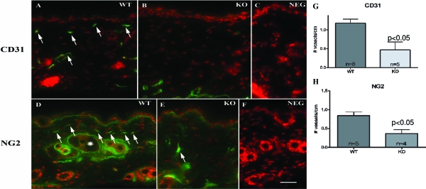
Figure 4.
Dermal microvasculature is reduced in epidermal vegf−/− mice. Frozen sections (5 μm) from epidermal vegf−/− versus WT biopsies (n = 3 each) were immunostained with a primary antibody against the endothelial cell marker, CD31 (A–C), or the pericyte marker, NG2 (D–F). C and F are negative controls from CD31 and NG2, respectively. Immunostained capillaries are indicated by arrows. NG2 also labels cells that encircle pilosebaceous structures (D, asterisk). The density of immunopositive vessels in the papillary dermis (-pilosebaceous structures) was quantitated in randomly-obtained, coded micrographs (G, CD31; H, NG2). A–F, Mag bar = 10 μm.

