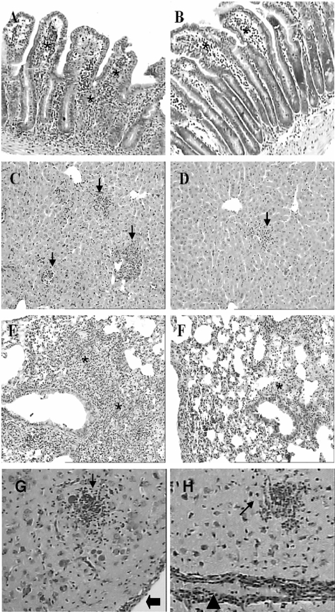
Figure 3.
Histological changes in the small intestine, liver, lung, and CNS of CCR2−/− and WT mice infected perorally with T. gondii. WT (A, C, E) and CCR2−/− (B, D, F) mice were inoculated with T. gondii and the peripheral organs were collected on day 8 after infection. Observed were inflammatory infiltrates in the lamina propria, epithelium, and submucosa (asterisks) in the small intestine (A, B); inflammatory foci (arrows) in the parenchyma of the liver (C, D); and inflammatory cell infiltration within the alveolar walls (asterisks) in the lung (E, F). The inflammatory reaction was milder in the peripheral organs of CCR2−/− (B, D, F) compared to WT mice (A, C, E). The CNS from WT (G) and CCR2−/− mice (H) was analyzed on day 23 after infection. The lesions characterized by glial nodules (small arrows), vascular cuffing (arrowheads) and inflammatory cells in the meninges (large arrows) were similar in both lineages of mice in this period of infection. Slides were stained with hematoxylin and eosin. Original magnification, ×10.

