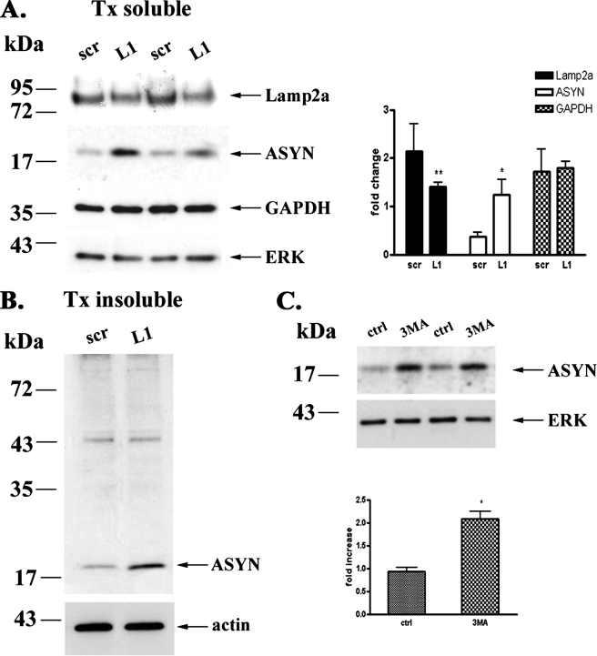
FIGURE 8.
Blockade of CMA or macroautophagy increases endogenous ASYN protein levels in postnatal ventral midbrain neurons. Three days after plating, postnatal ventral midbrain neurons (P1) were transduced with lentiviruses (m.o.i. 5) expressing Lamp2a (L1) or scr siRNA for 24 h. After 72 h, cells were assayed by Western blot for Lamp2a, ASYN, GAPDH, and ERK (loading control) levels. A, left, representative Western blots from three separate experiments are shown. Right, densitometric analysis of the levels of Lamp2a, ASYN, and GAPDH. B, detergent (Triton X-100)-insoluble pellets from the L1 or scr-treated samples were solubilized in SDS sample buffer and assayed by Western blot for ASYN and β-actin (loading control) levels. Representative Western blots from three separate experiments are shown. C, cultures were incubated with 3-MA (10 mm) for 24 h. Untreated cells were used as a control (ctrl). Cell lysates were assessed by Western immunoblotting for ASYN levels. Upper panel, representative immunoblot of ASYN. Bottom panel, quantification of endogenous ASYN levels after 3-MA addition, compared with control. All results are expressed as the ratio to OD values of the corresponding controls, and data are presented as mean ± S.E. of three independent experiments. (*, p < 0.05; **, p < 0.01, one way ANOVA followed by the Student-Newman-Keuls' test).

