FIGURE 6.
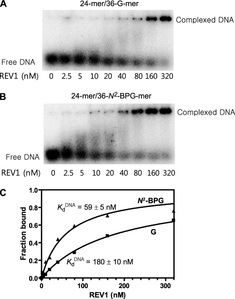
Estimation of the apparent KDNAd for REV1 to 24-mer/36-G-mer and 24-mer/36-N2-BPG-mer by electrophoretic mobility shift assay. A, 24-mer/36-G-mer; B, 24-mer/36-N2-BPG-mer. Reaction mixtures containing 0.5 nm of 32P-labeled 24-mer/36-mer primer-template duplex DNA were incubated with increasing concentrations of human REV1 (2.5–320 nm) and resolved on a 4% nondenaturing polyacrylamide gel to separate the free DNA and the REV1-DNA complexes. C, the fractions of REV1-bound DNA were plotted against the concentrations of free human REV1. Data were fit to a single-site binding equation, as described under “Experimental Procedures.” The fitted values of the apparent K DNAd are indicated in the figure. The following values of Kd were estimated: 24-mer/36-G-mer (▪), 180 ± 10 nm; 24-mer/36-N2-BPG-mer (▴), 59 ± 5 nm.

