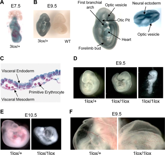
Figure 2. Essential role for Dot1L in mouse embryonic development.
(A) A representative X-gal stained 7.5-dpc Dot1L3lox/+ embryo demonstrating ubiquitous Dot1L transcription throughout the embryo. (B) A representative X-gal stained 9.5-dpc Dot1L3lox/+ embryo demonstrating ubiquitous Dot1L transcription throughout the embryo with elevated Dot1L expression in the indicated regions. (C) A representative X-gal stained 9.5-dpc Dot1L3lox/+ yolk sac demonstrating Dot1L transcription in visceral endoderm, visceral mesoderm, and primitive erythrocytes. (D) Representative pictures of 9.5-dpc Dot1L1lox/+ and Dot1L1lox/1lox embryos. Dot1L1lox/+ embryos (left) were indistinguishable from wild-type embryos. Most Dot1L1lox/1lox embryos were undersized, had an enlarged heart (cardiac dilation) and stunted tail (center), while approximately 15% exhibited developmental arrest at E8.5 (right). (E) Representative pictures of a 10.5-dpc Dot1L1lox/1lox embryo (right) and a heterozygous littermate (left). (F) Representative pictures showing the yolk sac vasculature of 9.5-dpc Dot1L1lox/+ (left) and Dot1L1lox/1lox (right) embryos. The vasculature of the Dot1L1lox/1lox yolk sac is thinner and less organized than that of the heterozygous littermate.

