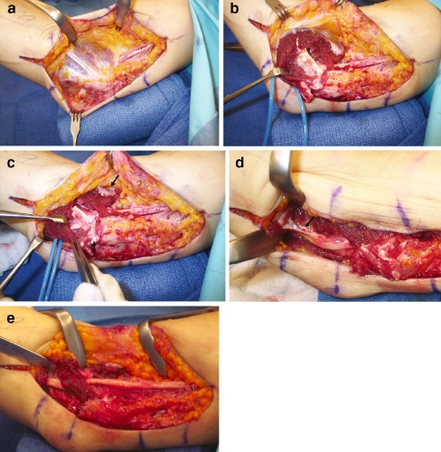
Figure 4.
(A) This patient has had a previous right Learmonth submuscular transposition. The ulnar nerve is seen proximally coursing under the pronotor flexor mass and anterior to the medial epicondyle in its transposed location. (B) The surgeon has retracted the soft flexor pronator muscle to identify the tendinous fascia (corresponding to “C” in Fig. 5B), which remains after the primary surgery. This intramuscular tendinous fascia is compressing the ulnar nerve. (C) The fascial flaps have been designed to be closed loosely over the ulnar nerve (arrows); however, they will not be needed as some muscle has been maintained above the ulnar nerve to serve as a method of not allowing the nerve to sublux back into the ulnar groove. The tendinous bands overlying the nerve will be removed and a limited neurolysis will be performed. (D) The tendinous fascia between the ulnar innervated flexor carpi ulnaris and the median innervated muscles is markedly kinking the nerve distally (arrow) and will be removed. This tendinous fascia would correspond to “B” in Fig. 5B. (E) At the conclusion of the procedure, the nerve lies in a straight location with no compression and no kinking of the nerve either proximally or distally.

