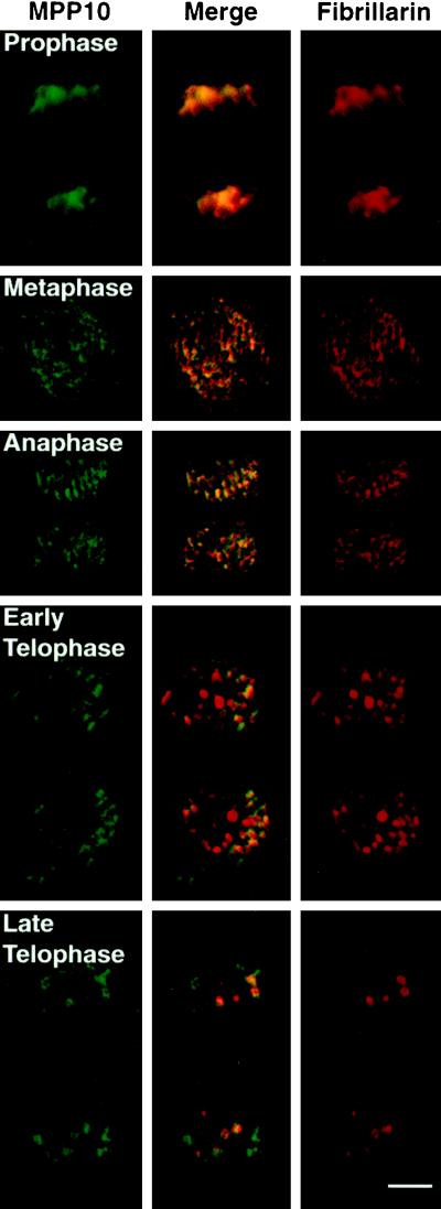
Figure 6.
Localization of MPP10 and fibrillarin during M phase of the cell cycle. Fixed HEp-2 cells were stained with affinity-purified guinea pig anti-MPP10 and mouse anti-fibrillarin, and staining was detected with fluorescein-labeled anti-guinea pig and rhodamine-labeled anti-mouse. Images were obtained by confocal microscopy. Bar, 5 μm.

