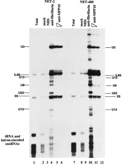
Figure 7.
Immunoprecipitation of U3 snoRNA by anti-MPP10. Protein A beads were incubated with no antibodies (mock), normal human serum (NHS), mouse monoclonal antibodies (72B9) to fibrillarin (anti-fibrillarin), or affinity-purified guinea pig anti-MPP10 (anti-MPP10; 10 μl, lanes 5 and 11; 2.5 μl, lanes 6 and 12). The resulting beads were washed and incubated with HeLa cell sonicates. RNA from the resulting immunoprecipitates was purified, end-labeled with 32P-labeled pCp, fractionated on 8% denaturing polyacrylamide gels, and autoradiographed. Lane 1 contains total RNA purified from 1% the number of cells used in the immunoprecipitations. The indicated positions of major RNAs were determined by comparison with the migration of pBR322 digested with MspI, end-labeled, and denatured. We do not consider the presence of 5S and 5.8S rRNAs in the precipitates to be specific because these RNAs are found in precipitates of splicing snRNPs also.

