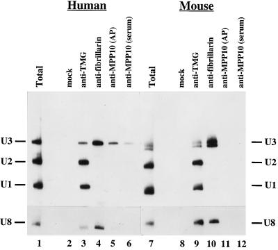
Figure 8.
Human and mouse anti-MPP10 immunoprecipitations analyzed by Northern blot analysis. Protein A beads were incubated with no antibodies (mock), mouse monoclonal antibodies to the TMG cap (anti-TMG), mouse monoclonal antibodies (72B9) to fibrillarin (anti-fibrillarin), affinity-purified guinea pig anti-MPP10 (anti-MPP10; affinity purified, AP), or anti-MPP10 guinea pig serum (anti-MPP10 [serum]), washed, and incubated with HeLa (lanes 2–6) or mouse L (lanes 8–12) cell sonicates. RNA from immunoprecipitates was isolated, fractionated on 8% denaturing polyacrylamide gels, blotted to Zeta-Probe, and hybridized with U1, U2, and U3, or U8 probes. Lanes 1 and 7 contain total RNA from 10% the number of cells used in the immunoprecipitations.

