
Figure 7.
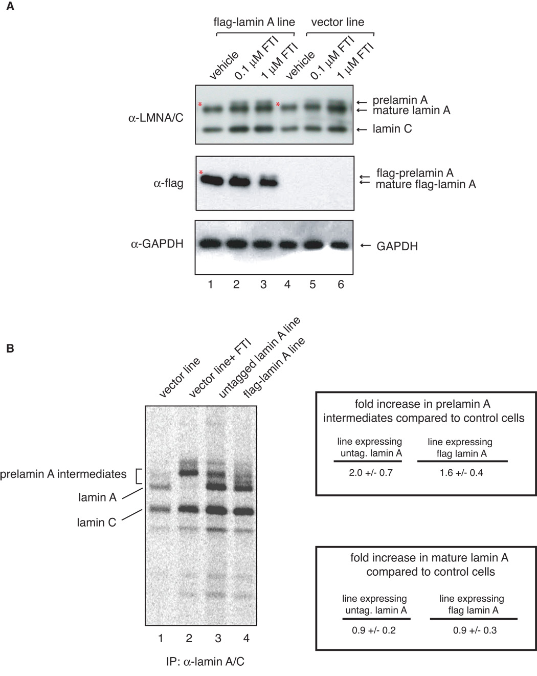
A There is no detectable accumulation of prelamin A in untreated flag-lamin A expressing fibroblasts. Fibroblasts expressing flag-lamin A and control at passage 10 were treated with vehicle only (lanes 1 and 4), and 0.1 µM (lanes 2 and 5) or 1 µM (lanes 3 and 6) farnesyl transferase inhibitor L-744832 for 48 hours, collected and analyzed by western blot analysis with lamin A/C (top panel), flag (middle panel) and GAPDH (lower panel) antibodies. To the best of resolution we do not see prelamin A accumulation in vector and flag-lamin A expressing fibroblasts treated with vehicle, even at lighter exposure (data not shown). However, Lamin A and flag-lamin A accumulate in FTI-treated control and flag-lamin A expressing fibroblasts. Asterisks show absence of prelamin A and flag-prelamin A in vehicle-treated cells. B. Immunoprecipitation analysis of prelamin A intermediates in metabolically labeled cells. Control fibroblasts, which were either untreated (lane 1) or treated with FTI (lane 2), and fibroblasts expressing either untagged (lane 3) or flag-tagged (lane 4) human wild type lamin A and were subjected to long term metabolic labeling and analyzed by fluorography as described in Materials and Methods. Biosynthesis of lamin A precursors and mature lamin A was visualized by phosphorimager and quantitated. Amounts of prelamin A intermediates and mature lamin A in each sample were normalized to lamin C and are shown as fold increase over that of control cells.