
FIGURE 1. Silencing of endogenous hSPRY2 decreases the anti-apoptotic actions of serum.
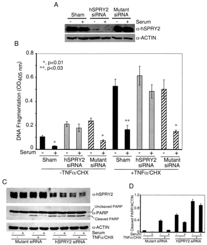
Panel A, SW13 cells were transfected with sham and 20 nM each hSPRY2 siRNA or mutant hSPRY2 siRNA (Mutant siRNA) and 48 h later plated on 35-mm dishes. After 36 h of serum deprivation, cells were lysed in Laemmli buffer, and proteins were separated on 10% polyacrylamide gels. The amount of SPRY2 was detected by immunoblotting to assess the efficiency of silencing by the siRNA. Actin was used to assess equal loading of samples. Panel B, cells treated as described in panel A were plated after 48 h of transfection onto 24-well plates. After 36 h of serum deprivation, the cells were treated with or without serum for 15 min. Vehicle or a mixture of TNF-α (20 ng/ml) and CHX (25 μg/ml) were added to the cells and incubated for 1 h before measuring DNA fragmentation as described under “Experimental Procedures.” Data are the mean ± S.D. of A405 nm per 50,000 cells (n = 6). The significance of differences was assessed by Student’s unpaired t test. Panel C, same as panel B, except that cells were lysed in Laemmli sample buffer, and proteins were separated by SDS-PAGE followed by immunoblotting with cleaved and total PARP antibodies. Panel D, quantification of the cleaved PARP normalized to endogenous actin from three independent blots similar to panel C.