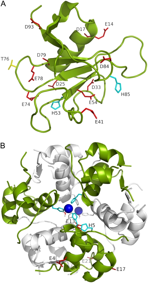
FIGURE 1.
(A) Structure of RNase Sa. Eleven carboxyl side chains, two histidines, and residue T76 are labeled and displayed as red, cyan, and yellow sticks, respectively. The N-terminal residue happens to be an aspartate. The side chain of this residue was not titrated in the constant-pH simulations, and is not shown here. (B) Structure of zinc-insulin hexamer. The two trimers are shown in the foreground and background in green and gray, respectively. The two zinc ions and the coordinating histidines and water molecules are shown. In addition, the five titrated side chains of one monomer are labeled and shown as sticks.

