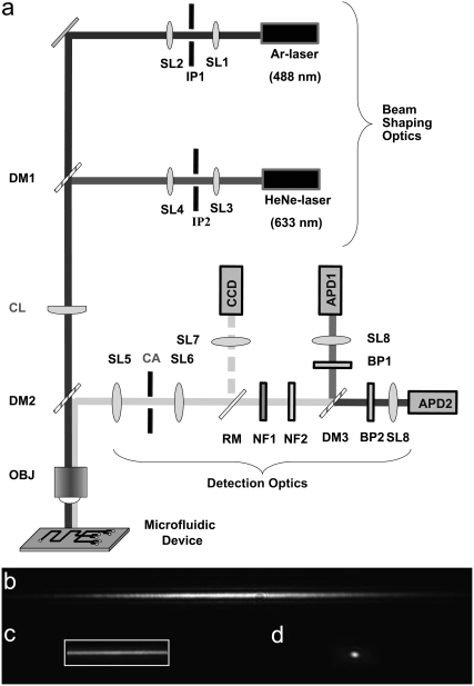
FIGURE 1.
(a) Schematic diagram of the key optical components in the CICS system. Reflected images of the illumination volume in (b) CICS with no aperture, (c) CICS after the 620 × 115 μm rectangular aperture, and (d) standard SMD with no pinhole. The standard SMD illumination volume resembles a football that extends in and out of the plane of the page whereas the CICS observation volume resembles an elongated sheet or plane that also extends in and out of the page. The CICS observation volume is expanded in 1-D using a cylindrical lens (CL) and then filtered using a rectangular aperture (CA). In the absence of a confocal aperture in b, the CICS illumination profile is roughly Gaussian in shape along the x, y, and z axis, chosen to align with the width, length, and height of a microchannel, respectively. The addition of the confocal aperture in c, depicted as a rectangular outline, allows collection of fluorescence from only the uniform center section of the illumination volume. Abbreviations: APD, avalanche photodiode; BP, bandpass filter; CA, confocal aperture; CCD, CCD camera; CL, cylindrical lens; DM, dichroic mirror; IP, illumination pinhole; NF, notch filter; OBJ, objective; RM, removable mirror; and SL, spherical lens.

