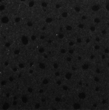
FIGURE 7.
A fluorescence confocal microscopy image of BLES-albumin (1:4 w/w, with 1% FITC-labeled BSA) films. Scan area is 50 × 50 μm. Films were prepared by the same procedures as in Fig. 1. The LB films were deposited onto glass coverslips at 30 mN/m. This image demonstrates that albumin coexists with BLES within the LE phase of the monolayer at a π higher than the πe of albumin (i.e., >20 mN/m).

