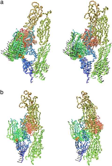
FIGURE 3.
Displacement vectors of Cα atoms of integrin in the lowest- (a) and third-lowest- (b) frequency normal modes of model B (see text) drawn in stereo with the backbone. Leu375, Leu389, and Arg633 are shown by a space-filling model. The images were generated with a VMD program (62).

