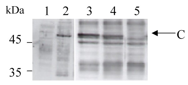
Figure 2.
Expression and display of calreticulin in Sf9 cells infected by the cDNA library. Crude cell extracts (10 μg of proteins) from non-infected Sf9 cells (lane 1), infected cells before panning (lane 2, 3), positive sorted cells after panning (lane 4) or negative sorted cells after panning (lane 5) were run on a SDS-PAGE (12%) and analysed by Western Blot against maize (lane 1, 2) or tobacco anti-calreticulin antiserum, (lane 3, 4,5). Position of size marker is indicated on the left. Arrow points to calreticulin (C) band.

